Volume 3 : Issue 3, September 2013

Table of Contents,
Issue 1 (11 Sept 2013), pp.68-88
|
Research Title/ |
Article Information/ |
Download |
|
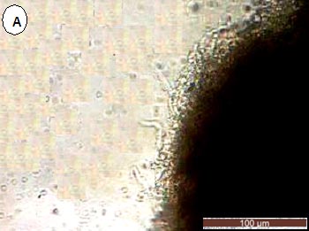
Development and Biological Characteristics of Brown Quail (Coturnix ypsilophora) Embryonic Fibroblast Primary Cells
|
Original Article, C13 ABSTRACT: Using tissue explantation and cryopreservation biotechniques, a quail embryonic fibroblast primary cell line was successfully developed, which performed by using 20 quail embryo samples and a stock of 25 cryovials, each one containing 3.0×10⁶ cells. Most of the cells were apparently fibroblasts in their morphology, and the population doubling time (PDT) was about 48 h. The cells were tested for microbial contamination and found free of infections from bacteria, fungi, viruses and mycoplasms. The total chromosome number of a diploid cell was 78 According to karyotyping and chromosome analysis. All the tests showed that the quality of the cell line conforms to the quality criteria of the ATCC (American type culture collection).This work succeeded not only in preserving the genetic resources of quail cells, but it also established a new protocol to preserve cell of avian breeds. |
|
|
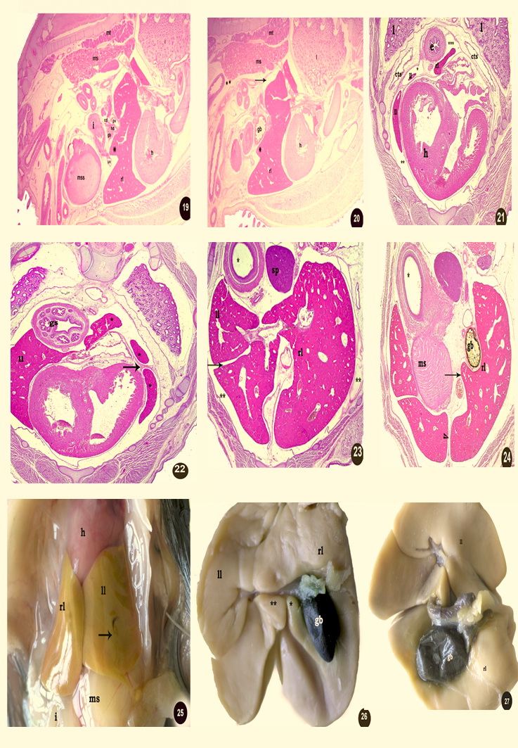
Dynamics of Liver Development in Dandarawi Chicken
|
Original Article, C14 ABSTRACT: The present work was carried out on 50 chick embryo of Dandarawi chicken collected from Assiut University Farm at a 3, 7, 9, 11, 13, 15, 17 and 19 day of prehatching life. At the 3rd day of incubation, the hepatic diverticulum gave dorsal and ventral parts in relation to the ductus venosus. At the 7th day, the dorsal and ventral parts became the left and right lobes respectively where the gall bladder was located on visceral surface of the right lobe and a transverse fissure dividing the left lobe into dorsal and ventral parts. Also, the vitelline veins caudal to the liver anastomosed together forming the portal vein, which gave off left portal branch to the left lobe of liver and continued as right portal branch to the right lobe. At the 9th day, the right lobe was longer and higher than the left one where the right lobe was in contact dorsally with the mesonephros and the left one was separated from the mesonephros by the glandular stomach. At the 11th day, the interlobar fissure was occupied mainly by the umbilical vein. At the 13th day, the parietal surface of the two lobes was related to the heart and body wall and the gall bladder increased in size and extended laterally. At the 15th day, the cranial end of the right lobe had three processes dorsal, middle and ventral but the cranial end of the left lobe had two processes dorsal and ventral. The duct system of the right lobe was hepatocystic and cystoenteric, but that of the left was hepatoenteric duct. |
|
|
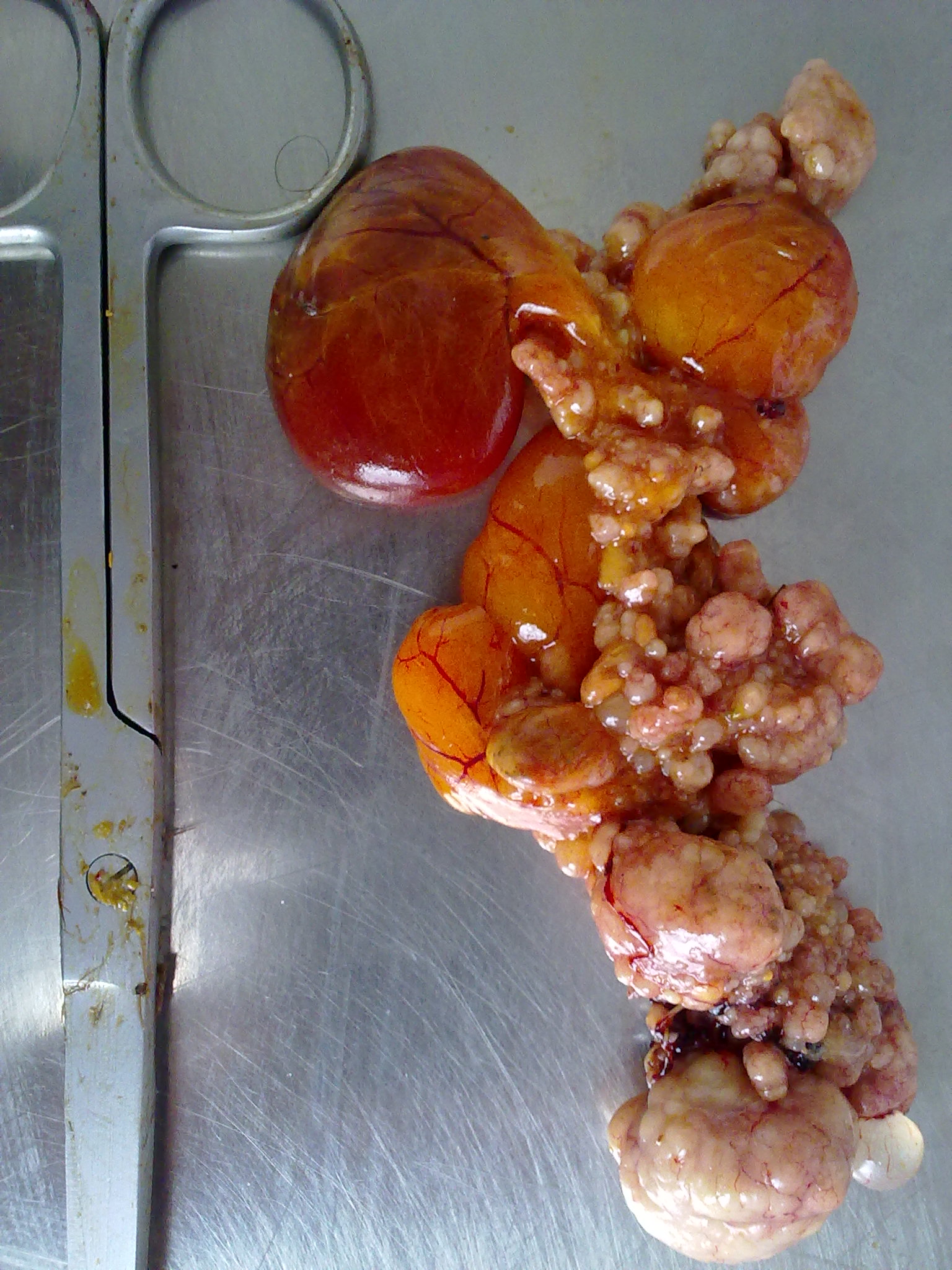
Intestinal Metastasis of Ovarian Adenocarcinoma in a Hen (Gallus domesticus) |
Original Article, C15 ABSTRACT: An aged dead adult native hen was referred for Necropsy. Grossly, pedunculated, firm, greyish-white fleshy growths were found attached to the serosal surface of ovary together with spread over the whole of intestine serosa. Microscopically, the ovarian growths consisted of a tubular pattern confirmed as adenocarcinoma with metastasis on the intestines. Key words: Intestine, metastasis, ovarian adenocarcinoma and pathological findings |
|
|
Comparison between Haemagglutination Inhibition Test and Enzyme Linked Immune Sorbent Assay in Evaluation of Newcastle disease Antibodies in Japanese Quails
|
Original Article, C16 ABSTRACT: Comparison between Haemagglutination Inhibition (HI) test and Enzyme Linked Immune Sorbent Assay (ELISA) in evaluation of humoral immune response to Newcastle disease (ND) in vaccinated quails was investigated. The obtained results showed higher NDV antibody titer (2, 5 and 8) log2 at zero, 14 and 28 day post vaccination in serum of La Sota vaccinated quails when the antigen used in HI test was homologous (La Sota), whereas lower antibody response (2, 4 and 6) log2 were obtained when the antigen used was heterologous (Hitchner B1). However, antibody titers in serum sample from Hitchner B1 vaccinated quails were (2, 4 and 6) log2 at zero, 14 and 28 day post vaccination against both homologous (Hitchner B1) and heterologous (La Sota) antigens. Non treated sera from control non vaccinated quails gave false positive results (6 log2) in HI test. Treatment of serum by heat (56´C /30 minutes) or by washing with 10% Chicken red blood overcome false positive results obtained by non treated serum in HI test. No difference between chicken and quail red blood cells were observed when used as red blood cells indicators in HI test to detect Newcastle disease virus antibodies of vaccinated quails. Two commercial ELISA kits used in this study failed to detect any positive sera (0.0 %) for ND antibodies in both vaccinated and non vaccinated quails at different time periods. |
|